 The aim of the Molecular Interactions Facility is to provide scientific guidance, support, and access to biophysical instrumentation for the quantitative characterization of molecules (size, shape & molar mass) and their interactions (stoichiometry & affinity).
In 2009 this Facility obtained the ISO9001 certificate according to the AENOR code ER-0286/2009. In October 2024, the Facility became a member of the network of Laboratories and Scientific and Technical Infrastructures of the Community of Madrid (REDLAB), with the assigned registration number RLAB-036.
SERVICES OFFERED:
Analytical ultracentrifugation (AUC): Determination of the size, degree of homogeneity, proportion of species, stoichiometry and affinity of proteins and other biological macromolecules in solution by Sedimentation Velocity and Equilibrium.
Dynamic Light Scattering (DLS): Determination of the degree of homogeneity of macromolecular complexes in solution, measurement of their translational diffusion coefficient and hydrodynamic radius.
Analytical Size Exclusion Chromatography coupled to Multi-Angle Light Scattering (SEC-MALS): Separation of the different macromolecular species present in the sample according to their size and determination of their molecular weight.
Biolayer Interferometry (BLI):Measurement of binding affinities and rates of association and dissociation within interactions between proteins, peptides, nucleic acids, small molecules, and lipids in real.
Fluorescence Correlation Spectroscopy (FCS): Study of protein polymerization and aggregation and measurement of molecular interactions rendering complexes.
Fluorescence Intensity and Anisotropy/Polarization: Detection and quantification of molecular interactions such as those involving proteins, nucleic acids, peptides or ligands, in the picomolar range.
For more information, contact with:
- Dr. Carlos Alfonso (carlosa@cib.csic.es). Phone: 91 837 3112 ext. 442711.
- Dr. Juan Román Luque (luque@cib.csic.es). Phone: 91 837 3112 ext. 442697
- Facility e-mail address: uanalitica@cib.csic.es. Phone: 91 837 3112 ext. 442697.
FACILITY EQUIPMENT
- Analytical ultracentrifugation (AUC): Two Beckman-Coulter analytical ultracentrifuges, an Optima XLA equipped with UV-VIS absorbance optics and an Optima XLI with integrated UV - VIS and Raleigh interference optics.
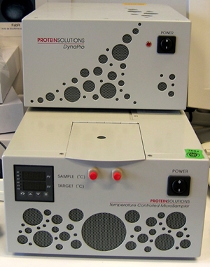
- Dynamic Light Scattering (DLS): A DynaPro MS/X model instrument from Wyatt Inc.
- Analytical Size Exclusion Chromatography coupled to Multi-Angle Light Scattering (SEC-MALS): A multi-angle laser light scattering detector equipped with a flow cell (DAWN EOS model, Wyatt Inc.) connected to an Optilab rEX refractive index flow detector from Wyatt Inc.
- Biolayer Interferometry (BLI): A ForteBioBLItzTM interferometer.
- Fluorescence Correlation Spectroscopy (FCS): A MicroTime 200 (PicoQuant) time-resolved confocal fluorescence microscope with single molecule sensitivity, picosecond pulsed (481 + 635) nm excitation, PicoHarp 300, 3D scanning and four channel detection (with two MPD and two AQR-SPADs).
- Fluorescence Intensity and Anisotropy/Polarization Spark® Multimode microplate reader (Tecan) equipped with polarizers for anisotropy measurements and with filters for wavelength selection [Ex: 360 (35), 485 (20), 530 (25), 560 (20), 620 (20). Em: 340 (20), 465 (35), 535 (25), 595 (35), 620 (20), 680 (30)].
Instructions
Users who wish to make a reservation or request more information about the techniques offered must contact the Facility by email (uanalitica@cib.csic.es) or telephone (918373112 Ext. 442697).
Sample and buffer requirements for Analytical Ultracentrifugation assays can be consulted here.
Rates, that depend upon the group’s institution (CSIC, universities, private centers, companies), include the following:
- Experimental design.
- Data acquisition setup.
- Assembly/disassembly and cleaning of cells.
- Raw data supplied as computer readable text files.
- A brief report with information about the quality of the sample (i.e., size heterogeneity, degree of association, etc.) and a preliminary gross estimate of cell-average molar masses and sedimentation coefficients. Delivery deadlines for sedimentation velocity and sedimentation equilibrium reports will be 3 and 5 working days, respectively.
Detailed analysis and interpretation of the results and subsequent preparation of figures and text for publication are not part of the fee-based service provided by this facility, but rather fall into the category of collaborative research, the conditions of which must be negotiated in advance between the principal investigator of the user’s work group and the staff of the Facility.
Members
>
Person in Charge
| Oscar Mariano Nuero García |
| Juan Roman Luque Ortega |
More Info
Analytical Ultracentrifugation (AUC) is an important group of powerful methods (sedimentation equilibrium and velocity) which allow determining the size, the approximate overall shape and the degree of homogeneity of proteins and other biological macromolecules in solution. These methods are specially adapted for the detection and quantitative analysis (proportion of species, stoichiometry, affinity, reversibility) of interactions leading to the formation of macromolecular complexes, including protein-protein, DNA-protein, and receptor-ligand interactions [Howlett et al. (2006) Curr. Opin. Chem. Biol. 10:430-436; Lebowitz et al. (2002) Protein Sci. 11:2067-2079; Minton (2000) Exp. Mol. Med. 32:1-5; Rivas et al. (1999) Methods 19:194-212].


Since its foundation in 1994, this CIB Facility has given scientific and technical support in the biophysical characterization of proteins and their interactions to a large number of research groups in the CIB, CSIC other centres, universities and other centres of domestic and foreign research. Moreover, the Facility counts with the advice and collaboration of international experts in the field and has participated in more than 300 publications since 1994.
Dynamic Light Scattering (DLS) provides complementary information to AUC, with two main advantages, relative to other commonly used analytical methods: low sample volume requirements (15-45 µl) and quick delivery of results (<10 min).
DLS allows determining the degree of homogeneity of macromolecular complexes in solution, to measure the translational diffusion coefficient of macromolecules and to calculate their hydrodynamic radius. This instrument (DynaPro MS/X model, Wyatt Inc.) is very well designed to study proteins and macromolecular assemblies (including polymers and liposomes), has a very easy operation (cuvette) and a Peltier temperature regulation system (4-60°C) and can be used to analyze different types of macromolecules such as proteins, polysaccharides, nanoparticles and lipid micelles.

Analytical Size Exclusion Chromatography coupled to Multi-Angle Light Scattering (SEC-MALS) separates the different macromolecular species present in the sample according to their size, and measures simultaneously their concentration and the intensity of the light scattered by these species, allowing the obtaining of their molecular weight.

For the rapid detection and real-time determination of the kinetic and equilibrium properties of homo- and hetero-associations of biological macromolecules and complexes in solution is also possible to use this instrument with a Composition Gradient (CG-MALS). This technique is based upon the methods of static light scattering recently developed by the group of Dr. Allen Minton, NIH [Attri & Minton (2005) Anal. Biochem. 337:103-110]. Main module is a multi-angle laser light scattering detector equipped with a flow cell (DAWN EOS model, Wyatt Inc.) that is connected to a refractive index flow detector and to a triple-syringe programmable pump. The information is acquired in minutes (important when the stability of the sample is a factor to consider). Moreover, the system allows determine the association/dissociation kinetics of a given macromolecular reaction (providing that the relaxation time of the reaction is in the minute range).
The Facility has carried out several studies on the oligomerization and polymerization of proteins by the combination of ultracentrifugation and light scattering techniques (Monterroso et al. (2013) Methods 59: 349–362; del Castillo et al. (2011) Biochemistry, 50: 1991–2003).
Biolayer Interferometry (BLI) is a simple, optical dip-and-read assay system useful for measuring interactions between proteins, peptides, nucleic acids, small molecules, and lipids in real time and small sample volumes (4.5 µL). BLI provides direct binding affinities and rates of association and dissociation between a molecule, immobilized on a fiber-optic biosensor, and the analyte in solution. Since it requires nanomole amounts of sample, it is an excellent choice for the study of proteins that are challenging to isolate. BLI also has the advantage of quantifying protein concentration from heterogeneous crude lysates, since refractive index changes in the medium do not affect shifts in the interference pattern


Fluorescence Correlation Spectroscopy (FCS) allows evaluating the temporal fluctuations of the fluorescence intensity to obtain insight into the mobility of the molecules, which is related with their size and shape. In this way, protein polymerization and aggregation can be studied. FCS is also a powerful tool for the detection and measurement of molecular interactions rendering complexes substantially bigger than the free species. FCS constitutes an ideal complement for other methodologies available in the facility, such as analytical ultracentrifugation and light scattering (Monterroso et al. (2013) Methods 59: 349–362).


Fluorescence Intensity and Anisotropy/Polarization measurements allow the detection and quantification of molecular interactions such as those involving proteins, nucleic acids, peptides or ligands with high sensitivity and low sample consumption. Anisotropy is particularly suitable for this purpose, as it depends on molecular volume and hence, it is sensitive to changes in size that occur upon complexation. The high sensitivity of these measurements (picomolar) enables evaluation of high affinity interactions under true equilibrium conditions. By using a plate reader, measurements are fast and can be performed simultaneously on hundreds of samples of low volume (a few microliters), opening the possibility of high throughput screening applications of interest in biotechnology and biomedicine.
